eyeball

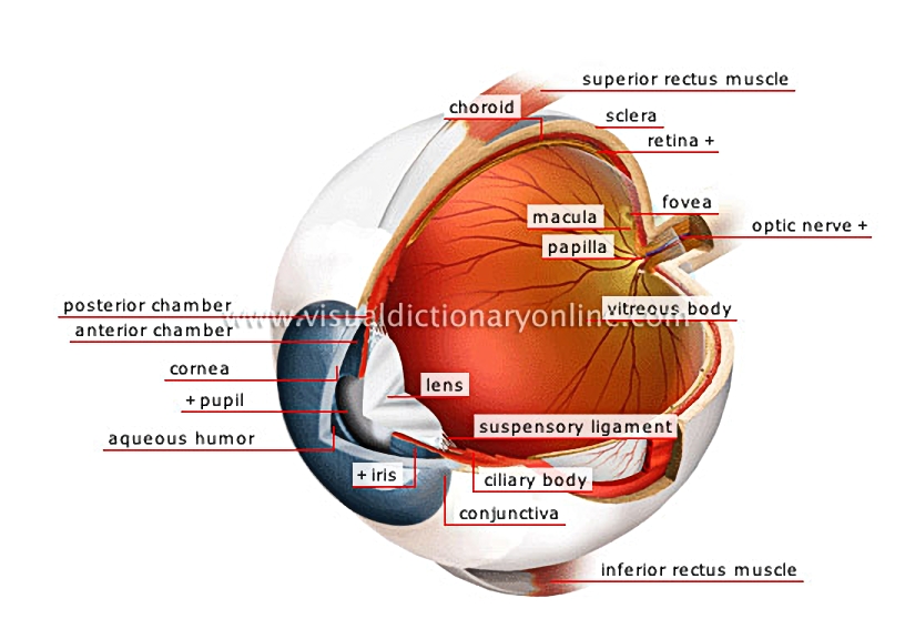
Enclosed in a bony cavity (orbit) and moved by six muscles, this complex organ collects light signals and transmits them to the brain to form images.
eyeball 						Strong fibrous opaque membrane covered by the conjunctiva; it surrounds the eyeball and protects the inner structures.				  							Muscle allowing the eyeball to move upward.				  							Central depression of the yellow spot composed entirely of cones; the place where visual acuity is at its maximum.				  							Transparent elastic area of the eye; focuses images on the retina to obtain clear vision.				  							Richly veined membrane located between the sclera and the retina, to which it carries nutrients and oxygen.				  							Inner membrane at the back of the eye covered in light-sensitive nerve cells (photoreceptors); these transform light into an electrical impulse that is carried to the optic nerve.												Area of the retina where the cones are concentrated; it plays an essential role in day vision and the perception of colors.				  							Nerve formed by the juncture of the nerve fibers of the retina; it carries visual information to the brain, where it is interpreted.				  							Protuberance formed by the anterior terminal part of the optic nerve in the retina; it has no light-sensitive cells and thus no vision. It is also called the blind spot.				  							Transparent gelatinous mass (almost 90% of the eye); it maintains constant intraocular pressure so the eye keeps its shape.				  							Muscle allowing the eyeball to move downward.				  							Muscle tissue secreting the aqueous humor; its muscles enable the lens to change shape to adapt vision for near or far.				  							Fibrous tissue connecting the ciliary body to the lens, holding it in place inside the eyeball.				  							Colored central portion of the eyeball composed of muscles whose dilation or contraction controls the opening of the pupil.												Fine transparent mucous covering the sclera and inner surface of the eyelid; it facilitates sliding thus giving the eyeball its wide range of movement.				  							Transparent liquid contained in the anterior and posterior chambers; it nourishes the iris and maintains the pressure and shape of the eye.				  							Central orifice of the eye whose opening varies to regulate the amount of light entering the eye; light causes the pupil to contract.												Transparent fibrous membrane extending the sclera and whose curved shape makes light rays converge toward the inside of the eye.				  							Cavity of the eye between the cornea and the iris containing the aqueous humor.				  							Cavity of the eye between the iris and the lens containing the aqueous humor.
See Also


photoreceptors

eye

contact lenses

eye protection

eye protection

sclera

Strong fibrous opaque membrane covered by the conjunctiva; it surrounds the eyeball and protects the inner structures.

superior rectus muscle

Muscle allowing the eyeball to move upward.

fovea

Central depression of the yellow spot composed entirely of cones; the place where visual acuity is at its maximum.

lens

Transparent elastic area of the eye; focuses images on the retina to obtain clear vision.

choroid

Richly veined membrane located between the sclera and the retina, to which it carries nutrients and oxygen.

retina

Inner membrane at the back of the eye covered in light-sensitive nerve cells (photoreceptors); these transform light into an electrical impulse that is carried to the optic nerve.
photoreceptors
photoreceptors

macula

Area of the retina where the cones are concentrated; it plays an essential role in day vision and the perception of colors.

optic nerve

Nerve formed by the juncture of the nerve fibers of the retina; it carries visual information to the brain, where it is interpreted.

papilla

Protuberance formed by the anterior terminal part of the optic nerve in the retina; it has no light-sensitive cells and thus no vision. It is also called the blind spot.

vitreous body

Transparent gelatinous mass (almost 90% of the eye); it maintains constant intraocular pressure so the eye keeps its shape.

inferior rectus muscle

Muscle allowing the eyeball to move downward.

ciliary body

Muscle tissue secreting the aqueous humor; its muscles enable the lens to change shape to adapt vision for near or far.

suspensory ligament

Fibrous tissue connecting the ciliary body to the lens, holding it in place inside the eyeball.

iris

Colored central portion of the eyeball composed of muscles whose dilation or contraction controls the opening of the pupil.
eye
eye

conjunctiva

Fine transparent mucous covering the sclera and inner surface of the eyelid; it facilitates sliding thus giving the eyeball its wide range of movement.

aqueous humor

Transparent liquid contained in the anterior and posterior chambers; it nourishes the iris and maintains the pressure and shape of the eye.

pupil

Central orifice of the eye whose opening varies to regulate the amount of light entering the eye; light causes the pupil to contract.
eye
eye

cornea

Transparent fibrous membrane extending the sclera and whose curved shape makes light rays converge toward the inside of the eye.

anterior chamber

Cavity of the eye between the cornea and the iris containing the aqueous humor.

posterior chamber

Cavity of the eye between the iris and the lens containing the aqueous humor.